Understanding Precancerous Mouth Cancer – A Vital Guide 2024
- 257
- 0
- 0
The rise in precancerous mouth cancer conditions in the mouth today can be attributed to lifestyle habits like tobacco and alcohol use, alongside the increasing prevalence of HPV infection. Neglecting oral hygiene and sun exposure also contribute. Early detection is crucial as it prevents progression to oral cancer, improving treatment outcomes and preserving quality of life. This underscores the importance of regular dental check-ups, lifestyle modifications, and public health initiatives to combat oral cancer.
Introduction
Mouth cancer, also known as oral cavity cancer, encompasses cancers that develop in various parts of the mouth, including the lips, gums, tongue, inner lining of the cheeks, roof, and floor of the mouth. It is categorized under head and neck cancers and shares similar treatment approaches with other cancers in this category. Identifying issues early and swiftly addressing them is essential for enhancing results and minimizing the chance of complications.
Types of Precancerous Mouth Cancer
Precancerous conditions in the mouth are cell changes increasing cancer risk. While not cancer, untreated abnormalities may progress to oral cancer. Common types include leukoplakia (white patches) and erythroplakia (red patches). Early detection and treatment are vital for preventing cancer development. Regular dental check-ups can help identify and manage these conditions, reducing the risk of oral cancer.

Two types of precancerous conditions in the mouth include:
- Leukoplakia: Leukoplakia presents as white or gray patches on the tongue, gums, inner cheeks, or roof of the mouth. While not always cancerous, leukoplakia lesions have the potential to progress to oral cancer if left untreated.
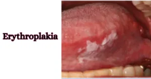
- Erythroplakia: Erythroplakia is characterized by abnormal red areas or spots on the mucous membrane lining the mouth, often without an identifiable cause. Erythroplakia lesions carry a higher risk of developing into squamous cell carcinoma compared to leukoplakia lesions.
Leukoplakia
Leukoplakia is known as white or grey patches on the tongue, inside cheeks, gums, or mouth floor. While not always cancerous, risk depends on cell abnormality (dysplasia) compared to normal cells. Healthcare teams monitor leukoplakia cases for signs of cancer, assessing cell shape, size, and appearance to gauge risk and guide treatment. Regular observation and timely intervention are crucial for preventing potential progression to oral cancer, emphasizing the importance of proactive dental care and monitoring for individuals with leukoplakia.

Risk Factors:
The four main risk factors for precancerous mouth cancer conditions of the mouth are:
- Tobacco use: Smoking cigarettes, cigars, or pipes, as well as using smokeless tobacco, significantly increases the risk of developing precancerous lesions in the mouth.
- Alcohol consumption: Heavy alcohol consumption, particularly when combined with tobacco use, escalates the risk of precancerous changes in the oral cavity.
- HPV infection: Certain strains of the human papillomavirus (HPV), especially HPV-16 and HPV-18, have been linked to an increased risk of developing oral precancerous lesions.
- Poor oral hygiene: Neglecting oral hygiene practices, such as regular brushing and flossing, can contribute to the development of oral precancerous conditions.
These factors play significant roles in the development of precancerous mouth cancer conditions in the mouth and are important considerations for prevention and early detection.
Signs & Symptoms:
Signs and symptoms of precancerous conditions in the mouth are:
- White or red patches: Abnormal patches of coloration on the tongue, gums, cheeks, or roof of the mouth.
- Persistent sores or ulcers: Non-healing sores or ulcers in the mouth that last for more than two weeks.
- Rough or thickened areas: Areas of the mouth that feel rough, thickened, or raised.
- Difficulty swallowing or chewing: Changes in swallowing or chewing habits, such as pain or discomfort while eating.
It’s important to note that these signs and symptoms can also be indicative of other oral health issues, so it’s essential to consult a healthcare professional for proper evaluation and diagnosis if experiencing any concerning symptoms. Early detection and intervention can significantly improve outcomes for individuals with precancerous mouth cancer conditions in the mouth.
Diagnosis:
The diagnosis of precancerous conditions in the mouth typically involves the following point:
- Clinical examination: A healthcare professional, often a dentist or oral surgeon, visually inspects the mouth, gums, tongue, and other oral tissues for any abnormal signs or symptoms.
- Biopsy: If suspicious lesions or areas are identified during the clinical examination, a biopsy may be performed.
- Imaging tests: In some cases, imaging tests such as X-rays, CT scans, or MRI scans may be ordered to assess the extent of tissue involvement and to evaluate for any potential spread of abnormal cells.
- HPV testing: For lesions suspected to be related to human papillomavirus (HPV) infection, testing for HPV DNA or RNA may be performed to confirm the presence of the virus.
- Histopathological evaluation: The biopsy sample obtained is examined by a pathologist under a microscope to assess the cellular characteristics and determine the presence of dysplasia or other precancerous changes.
These diagnostic steps help healthcare professionals accurately identify and characterize precancerous conditions in the mouth, guiding appropriate management and treatment decisions.
Treatment:
The treatment of precancerous conditions in the mouth typically involves the following approaches:
- Observation: In cases where the precancerous lesions are small and low-risk, healthcare professionals may choose to closely monitor the lesions over time without immediate intervention. Regular follow-up appointments are scheduled to assess any changes in the lesions and to determine if further treatment is necessary.
- Surgical removal: Precancerous lesions that are larger or higher risk may be surgically removed through procedures such as excision or laser surgery. This involves removing the abnormal tissue along with a margin of healthy tissue to ensure the complete removal of any potentially cancerous cells.
- Topical medications: In some cases, topical medications such as retinoids or topical chemotherapy agents may be applied directly to the precancerous lesions to help eliminate abnormal cells and reduce the risk of progression to cancer.
- Cryotherapy: Cryotherapy involves freezing the precancerous lesions using liquid nitrogen or another freezing agent. This destroys the abnormal tissue and promotes the growth of healthy new tissue.
- Photodynamic therapy: Photodynamic therapy (PDT) uses a combination of a light-sensitive drug and laser light to selectively target and destroy precancerous cells while minimizing damage to surrounding healthy tissue.
- Follow-up care: Regardless of the treatment approach used, regular follow-up appointments are essential to monitor for any recurrence of precancerous lesions or development of oral cancer. Lifestyle modifications, such as quitting smoking and reducing alcohol consumption, may also be recommended to reduce the risk of recurrence.
Individuals diagnosed with precancerous mouth cancer conditions in the mouth need to work closely with their healthcare team to determine the most appropriate treatment plan for their specific situation.
Erythroplakia
Erythroplakia is characterized by abnormal red areas or spots on the mouth’s mucous membrane, often without a clear cause. While not always cancerous, this precancerous condition poses a high risk of progressing to cancer. Approximately 50% of Erythroplakia lesions evolve into squamous cell carcinoma. Early detection and intervention are crucial to managing Erythroplakia and reducing the likelihood of cancer development.

Risk Factors:
Primary risk factors for erythroplakia include:
- Tobacco use: Smoking cigarettes, cigars, or pipes, as well as using smokeless tobacco, significantly increases the risk of developing erythroplakia.
- Alcohol consumption: Heavy alcohol consumption, particularly when combined with tobacco use, escalates the risk of erythroplakia formation.
- HPV infection: Certain strains of the human papillomavirus (HPV), particularly HPV-16 and HPV-18, have been linked to an increased risk of developing erythroplakia.
- Poor oral hygiene: Neglecting regular oral hygiene practices, such as brushing, flossing, and dental check-ups, can contribute to the development of erythroplakia lesions.
Signs & Symptoms:
- Abnormal red areas: Erythroplakia presents as abnormal red patches or spots on the mucous membrane lining the mouth, often with a velvety or granular appearance.
- No clear cause: Unlike normal redness or irritation, erythroplakia arises spontaneously without an obvious cause such as trauma or irritation.
- Persistent lesions: Erythroplakia lesions typically do not resolve on their own and persist for an extended period, lasting for weeks or months.
- High risk of cancer: While not always cancerous, erythroplakia carries a significant risk of developing into squamous cell carcinoma, with approximately 50% of lesions progressing to cancer if left untreated.
Diagnosis:
The diagnosis of erythroplakia involves the following four points:
- Clinical examination: A healthcare professional visually inspects the mouth, gums, tongue, and other oral tissues for any abnormal red areas or spots.
- Biopsy: Suspicious lesions are typically biopsied, where a small sample of tissue is removed from the affected area for laboratory analysis.
- Histopathological evaluation: The biopsy sample is examined by a pathologist under a microscope to assess the cellular characteristics and determine the presence of precancerous or cancerous changes.
- HPV testing (if indicated): In cases where HPV infection is suspected, testing for HPV DNA or RNA may be performed to confirm the presence of the virus, as certain strains of HPV are associated with an increased risk of erythroplakia.
Treatments:
Treatment of erythroplakia typically involves the following four approaches:
- Surgical removal: Excision or laser surgery may be performed to remove the erythroplakia lesions, along with a margin of healthy tissue to ensure complete removal of potentially cancerous cells.
- Photodynamic therapy (PDT): PDT involves using a light-sensitive drug and laser light to selectively target and destroy abnormal cells while minimizing damage to surrounding healthy tissue.
- Follow-up care: Regular follow-up appointments are essential to monitor for any recurrence of erythroplakia or the development of oral cancer.
- HPV vaccination: In cases where HPV infection is implicated in the development of erythroplakia, vaccination against HPV may be recommended to reduce the risk of recurrence and progression to cancer.
Things to Avoid in Precancerous Mouth Cancer
When dealing with precancerous mouth cancer conditions, it’s essential to avoid the following actions:
- Ignoring symptoms: Don’t ignore any signs or symptoms, such as white or red patches, persistent sores, or lumps in the mouth, as early detection is crucial for effective treatment.
- Delaying seeking medical advice: Don’t delay seeking medical advice if you notice any abnormal changes in your mouth. Prompt evaluation by a healthcare professional can lead to timely diagnosis and intervention.
- Neglecting oral hygiene: Don’t neglect regular oral hygiene practices, such as brushing, flossing, and routine dental check-ups, as maintaining good oral health can help prevent the development or progression of precancerous lesions.
- Continuing tobacco and alcohol use: Avoid tobacco use in any form, including smoking and smokeless tobacco, as well as excessive alcohol consumption, as these are significant risk factors for precancerous mouth conditions and oral cancer.
- Disregarding lifestyle modifications: Don’t disregard lifestyle modifications that can reduce the risk of precancerous mouth conditions, such as quitting smoking, reducing alcohol intake, eating a healthy diet rich in fruits and vegetables, and practicing sun safety to protect the lips from sun exposure.
Importance of Early Detection and Intervention

Early detection of precancerous conditions offers the best chance for successful treatment and prevents the development of oral cancer. Regular dental check-ups, maintaining good oral hygiene, avoiding tobacco and excessive alcohol consumption, and seeking medical attention for any concerning symptoms are essential for early detection and intervention.
It’s important to schedule an appointment with your doctor or dentist if you experience persistent signs and symptoms that last for more than two weeks and are concerning to you. While these symptoms may be indicative of various conditions, including infections, it’s essential to rule out more serious underlying causes, such as oral cancer. Early detection and intervention can significantly improve treatment outcomes and overall prognosis.
Conclusion
Precancerous conditions in the mouth may not always be visible or symptomatic, underscoring the importance of proactive dental care and regular screenings. By understanding the causes, symptoms, and significance of early detection, individuals can take proactive steps to preserve their oral health and reduce the risk of developing oral cancer. If you notice any abnormalities in your mouth, don’t hesitate to consult with a healthcare professional for evaluation and guidance.
FAQs
1. What are the common signs of precancerous mouth conditions?
Early signs include white or red patches, persistent sores, rough areas, and lumps in the mouth. These symptoms may indicate potential precancerous changes requiring medical evaluation.
2. What causes precancerous mouth conditions?
Risk factors include tobacco and alcohol use, HPV infection, poor oral hygiene, sun exposure, and dietary deficiencies. Identifying and avoiding these factors can help reduce the risk of developing precancerous lesions.
3. How are precancerous mouth conditions diagnosed?
Diagnosis typically involves a combination of clinical examination, biopsy, histopathological evaluation, and HPV testing if indicated. Early detection is crucial for timely intervention and prevention of progression to cancer.
4. Are precancerous mouth conditions reversible?
While not all precancerous lesions progress to cancer, early detection and intervention can prevent progression. Lifestyle modifications, regular dental check-ups, and avoiding risk factors are essential for managing and reducing the risk of precancerous conditions.
5. What is the importance of early detection and intervention?
Early detection allows for prompt treatment, improving outcomes and reducing the risk of complications. Regular screenings, maintaining good oral hygiene, and seeking medical evaluation for any concerning symptoms are key preventive measures.
Also Read:
6 Wellhealth Ayurvedic Health Tips for Prevention and Wellness
The 5 Benefits of Meditation for Students – 2024
References:
https://www.ncbi.nlm.nih.gov/pmc/articles/PMC7764090/
https://pubmed.ncbi.nlm.nih.gov/12139232/
https://www.ncbi.nlm.nih.gov/pmc/articles/PMC7515567/
Disclaimer: This guide provides general information on precancerous mouth cancer. It’s not a substitute for medical advice. Always consult healthcare professionals for diagnosis and treatment. We are not liable for personal health decisions.
